
Function of the Illuminator on Microscope: 5 Core Roles Explained
Function of the Illuminator on Microscope: 5 Core Roles Explained
Learn the exact function of the illuminator on microscope systems. Discover how properly controlled lighting reveals cellular structures, prevents optical distortion, and enhances image resolution.
Microscopy relies on two fundamental principles: magnification and illumination. While objective lenses handle magnification, understanding the primary function of the illuminator on microscope units is crucial for achieving clear, high-contrast specimen detail. Without a proper light source, even top-tier glass optics yield dark, uninformative views.
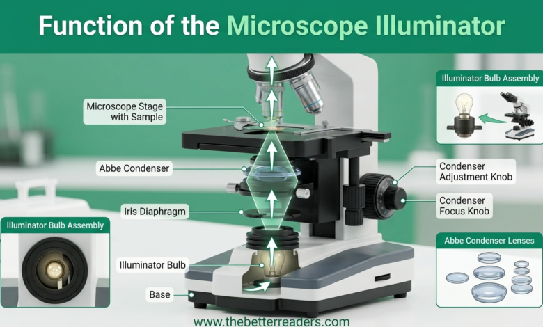
When examining biological samples, the primary function of the illuminator on microscope instruments is to generate photons that travel directly through the stage aperture. In this comprehensive study guide on thebetterreaders.com, we break down the structure, mechanics, and operational importance of the microscope illuminator, explaining how it works with condensers, diaphragms, and filter assemblies to deliver crisp optical views.
Short Answer: What Is the Function of the Illuminator on Microscope?
The primary function of the illuminator on microscope setups is to act as the built-in or external light source located at the base. It casts a direct, controllable, and uniform beam of light upward through the stage aperture to illuminate the mounted sample for detailed optical observation.
- Main Purpose: Provides uniform light needed to visualize transparent biological slides or solid samples.
- Control Features: Uses rheostat switches, blue daylight filters, and iris aperture adjustments to adjust light intensity.
- Optical Synergy: Feeds focused light directly into the condenser lens underneath the microscope stage.
1. The Crucial Role of the Illuminator in Microscopic Optics
Human vision requires reflected or transmitted light to register shape, color, and texture. In compound light microscopy, samples such as plant cells, bacterial smears, or blood tissue are extremely thin. The key function of the illuminator on microscope systems is to provide the energy required to pass through these samples and reach the observer’s eye.
Without understanding the core function of the illuminator on microscope hardware, laboratory users often struggle with dark images or severe glare. Modern illuminators allow precise brightness regulation to prevent oversaturation on delicate biological samples.
As objective magnification increases (from 4x up to 100x oil immersion), the field of view shrinks and the light pathway narrows. A high-output, adjustable function of the illuminator on microscope equipment is essential to maintain image brightness and contrast at higher magnifications.
2. Breakdown: 5 Core Functions of the Microscope Light Source
To understand how a compound microscope functions, let’s explore the five primary roles played by the light source unit:
- Directing Transmitted Light Rays: The primary function of the illuminator on microscope devices is shooting light rays upward through the condenser lens, specimen slide, objective lens, and eyepiece.
- Controlling Variable Light Intensity: Equipped with electronic rheostat dials, modern illuminators allow users to dim or brighten light output depending on sample density.
- Providing Even Field Distribution: Prevents uneven hot spots or dark borders, ensuring the entire specimen field receives uniform lighting.
- Supporting Specialized Lighting Techniques: Supplies continuous light required for darkfield, phase contrast, and polarized light microscopy modes.
- Enhancing Image Resolution and Color Fidelity: LED and daylight-filtered illuminators provide true color rendering without adding unwanted yellow hues.
3. Comparing Illuminator Technologies (LED vs. Halogen vs. Mirror)
Microscopes use various light technologies depending on their design, age, and intended laboratory application. Regardless of the technology used, the central function of the illuminator on microscope systems remains consistent—delivering dependable, bright light across the visual field.
| Illuminator Type | Heat Generation & Energy Efficiency | Best Suited Laboratory Application |
|---|---|---|
| LED (Light Emitting Diode) | Zero heat generated; highly energy efficient with long battery options. | Educational classrooms, living cell cultures, clinical diagnostics, and field work. |
| Tungsten-Halogen Bulb | Generates moderate heat; requires fan cooling in high-power setups. | Histology staining analysis, industrial inspection, and high-intensity research. |
| Adjustable Concave Mirror | No electrical consumption; relies entirely on external sunlight or room lights. | Vintage educational microscopes and field setups lacking electrical power. |
Explore more student learning resources on The Better Readers Main Hub. For additional business, technology, and media analyses, explore WeeklyMagazine Tech Section.
4. How the Illuminator Interacts with Other Optical Components
The illuminator does not work in isolation. It forms the start of a synchronized optical pathway where the main function of the illuminator on microscope systems is to feed light into adjacent components:
- Step 1: Emission from Illuminator: Light photons exit the LED or halogen housing at the base of the unit.
- Step 2: Aperture Control via Diaphragm: The iris diaphragm adjusts the width of the light column passing through.
- Step 3: Light Focusing via Condenser: The Abbe condenser focuses diffuse light rays into a tight cone directed at the glass slide.
- Step 4: Specimen Illumination: Light passes through the specimen on the stage, highlighting cellular details.
- Step 5: Objective Magnification: Objective lenses collect transmitted light and project a magnified image to the eyepiece.
5. Best Practices for Maintaining Microscope Illuminators
To extend bulb life and preserve the proper function of the illuminator on microscope devices during lab experiments, observe the following maintenance steps:
- Avoid Touching Bulb Glass with Bare Hands: Oil from fingertips creates hot spots on halogen glass bulbs, causing premature burning.
- Turn Down Voltage Before Shutting Off: Lower the rheostat dial before powering down to protect against voltage spikes when switching back on.
- Keep Lens Housing Free of Dust: Wipe the base collector lens periodically using specialized optical lens paper.
6. Frequently Asked Questions (FAQs & PAA)
Q1: What is the main function of the illuminator on a microscope?
The primary function of the illuminator is to supply steady, focused light that illuminates the specimen from below or above, allowing the objective lenses to produce a clear, sharp, magnified image.
Q2: What is the purpose of illumination in light microscopy?
Illumination creates visual contrast between microscopic structures and their background, enabling the observer to view cell walls, nuclei, and microorganisms clearly.
Q3: How do the condenser and illuminator work together on a microscope?
The illuminator acts as the light engine, while the condenser gathers and focuses those raw light rays into a precise light cone targeted directly at the specimen plane.
Q4: What are the main illuminating parts of a microscope?
The primary illuminating components include the illuminator (light bulb or mirror), the iris diaphragm, light intensity rheostat, and the sub-stage condenser lens assembly.
Q5: Is an LED illuminator better than a halogen bulb for a microscope?
LED illuminators are generally superior for modern routine microscopy because they produce daylight-balanced white light, generate zero heat, draw minimal power, and offer significantly longer operating lifespans.
7. Summary: The Foundation of High-Resolution Observation
Understanding the essential function of the illuminator on microscope systems is necessary for anyone using laboratory optics. By choosing the right lighting type and adjusting brightness appropriately, researchers and students can unlock clear, detailed, and high-contrast views across all biological specimens.
Syeda
Syeda is a dedicated SEO consultant, professional link builder, and author for thebetterreaders.com. She specializes in scientific educational guides, study materials, and search engine optimization for academic resources.